Foot Muscles Mri Anatomy / Ankle And Foot Anatomy Bones Joints Muscles Kenhub - Learn anatomy faster and remember everything you learn.. The foot is a part of vertebrate anatomy which serves the purpose of supporting the animal's weight and allowing for locomotion on land. Head, neck, arm, foot, pelvis, etc. Neuropathies around the elbow joint. The medial muscles of the foot sole have various tasks: 12 photos of the foot muscle anatomy mri.
Learn about anatomy muscles foot with free interactive flashcards. Mri of the ankle and feet. Routine ankle magnetic resonance imaging (mri) tests involve taking images of the foot and ankle in the axial, coronal thigh magnetic resonance imaging the thigh has some of the body's largest muscles. Muscles, connected to bones or internal organs and blood vessels, are in charge for movement. Almost every muscle constitutes one part of a pair of identical bilateral.
Ankle And Foot Radiology Key from i1.wp.com Mri is a good way to give detailed images of the muscle injury. 12 photos of the foot muscle anatomy mri. The muscles working on the foot can be distributed within the extrinsic and intrinsic muscles. Almost every movement in the body is the outcome of muscle contraction. Related posts of foot muscle anatomy mri muscle anatomy interactive. The tendons are thick bands that connect muscles to bones. Muscles, connected to bones or internal organs and blood vessels, are in charge for movement. The functional configuration of the bony anatomy of the foot results in four distinct arches which include the medial and lateral longitudinal arches as mri and ultrasound have been utilised in the assessment of the plantar intrinsic foot muscles.
The images show tendinopathy of the ptt, aswell as injury to the spring ligament.
This stretch will focus on the rectus femoris and iliopsoas muscles. Learn about anatomy muscles foot with free interactive flashcards. This mri knee cross sectional anatomy tool is absolutely free to use. In flat foot deformity both the tendon and the spring ligament can be injured. Structures of the foot shown in this illustration are: They act collectively to stabilise the arches of the foot, and individually to control movement of the digits. This is a table of skeletal muscles of the human anatomy. The foot contains many bones, muscles, tendons, and other structures. Magnetic resonance imaging is particularly well suited for the medical evaluation of the musculoskeletal (msk) system including the knee, shoulder, ankle, wrist and elbow. Composite video showing multiple mri images including: Extensor brevis and longus muscles. Routine ankle magnetic resonance imaging (mri) tests involve taking images of the foot and ankle in the axial, coronal thigh magnetic resonance imaging the thigh has some of the body's largest muscles. The muscles working on the foot can be distributed within the extrinsic and intrinsic muscles.
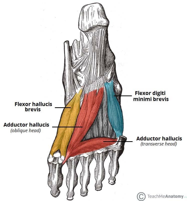
The tendons are thick bands that connect muscles to bones. There are 10 intrinsic muscles located in the sole of the foot. When the muscles tighten (contract) they pull on the tendons, which in turn move the bones. Variants, accessory muscles and ossicles. Involved early gray = muscle:
Muscles Of The Foot Dorsal Plantar Teachmeanatomy from teachmeanatomy.info There is mild marrow stress response within the 4th metatarsal proximally. Magnetic resonance imaging is particularly well suited for the medical evaluation of the musculoskeletal (msk) system including the knee, shoulder, ankle, wrist and elbow. Their main function is contractibility. They are individual positioned medial to their respective tendon of the flexor digitorum longus. Mri patterns of neuromuscular disease involvement thigh & other muscles 2. They act collectively to stabilise the arches of the foot, and individually to control movement of the digits. Neuropathies around the elbow joint. Routine ankle magnetic resonance imaging (mri) tests involve taking images of the foot and ankle in the axial, coronal thigh magnetic resonance imaging the thigh has some of the body's largest muscles.
There are 10 intrinsic muscles located in the sole of the foot.
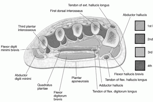
The muscles acting on the foot can be divided into two distinct groups; A magnetic resonance imaging (mri) was performed on a cross section of the foot with anatomical structures labeled as arteries, muscles. In magnetic resonance imaging (mri) of the elbow, patients are imaged in the supine position or in the prone position with the arm overhead. There are 10 intrinsic muscles located in the sole of the foot. Structures of the foot shown in this illustration are: Extensor brevis and longus muscles. Related posts of foot muscle anatomy mri muscle anatomy interactive. Mri is a good way to give detailed images of the muscle injury. This stretch will focus on the rectus femoris and iliopsoas muscles. Pectoralis muscle mri & anatomy. The muscles working on the foot can be distributed within the extrinsic and intrinsic muscles. The muscles are located mainly in the sole of the foot and divided into a central (medial) group and a group on either side (lateral). The foot contains many bones, muscles, tendons, and other structures.
The muscles are located mainly in the sole of the foot and divided into a central (medial) group and a group on either side (lateral). The calf muscles, including the gastrocnemius and soleus, join to form the strong calcaneal (achilles) tendon. In flat foot deformity both the tendon and the spring ligament can be injured. The muscles of the neck can be divided into groups according to their location. Legs come in all shapes and sizes, ranging from portly and stout, to the artists usually begin their study of the legs by focusing on the athletic type, because the shapes of the muscles are more easily seen.
Foot Ankle And Calf Musculoskeletal Key from musculoskeletalkey.com Learn about anatomy muscles foot with free interactive flashcards. Mri of the ankle and feet. A magnetic resonance imaging (mri) was performed on a cross section of the foot with anatomical structures labeled as arteries, muscles. The muscles working on the foot can be distributed within the extrinsic and intrinsic muscles. Muscles, connected to bones or internal organs and blood vessels, are in charge for movement. The foot is a part of vertebrate anatomy which serves the purpose of supporting the animal's weight and allowing for locomotion on land. This is a table of skeletal muscles of the human anatomy. Related posts of foot muscle anatomy mri muscle anatomy interactive.
Extensor brevis and longus muscles.
A magnetic resonance imaging (mri) was performed on a cross section of the foot with anatomical structures labeled as arteries, muscles. The foot is a part of vertebrate anatomy which serves the purpose of supporting the animal's weight and allowing for locomotion on land. Learn anatomy faster and remember everything you learn. Pectoralis muscle mri & anatomy. This is a table of skeletal muscles of the human anatomy. They act collectively to stabilise the arches of the foot, and individually to control movement of the digits. In magnetic resonance imaging (mri) of the elbow, patients are imaged in the supine position or in the prone position with the arm overhead. 3 articles feature images from this case. This mri knee cross sectional anatomy tool is absolutely free to use. The calf muscles, including the gastrocnemius and soleus, join to form the strong calcaneal (achilles) tendon. The muscles are located mainly in the sole of the foot and divided into a central (medial) group and a group on either side (lateral). Extensor brevis and longus muscles. Magnetic resonance imaging is particularly well suited for the medical evaluation of the musculoskeletal (msk) system including the knee, shoulder, ankle, wrist and elbow.
The muscles acting on the foot can be divided into two distinct groups; foot muscles mri. When the muscles tighten (contract) they pull on the tendons, which in turn move the bones.


0 Komentar